

Raman Microscopy Laboratory — Equipment
We are a research and service division of:
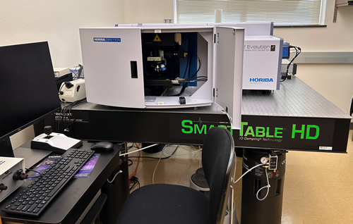
Our confocal Raman microscope was installed in 2022 and is supported through an NSF-MRI grant. The instrument is a Horiba LabRAM HR Evolution confocal Raman Microscope with a 532 nm (green) laser and a 266 nm (UV) laser, which focuses the laser beam on to the sample using a set of internal mirrors. It is also coupled with an Olympus microscope and an automated YZY-stage, for easy navigation of samples. While the elastic scattering is filtered out (Rayleigh line), the inelastic scattering is collected onto a detector/spectrometer, providing detailed information about the physical and chemical properties of the analyzed molecules. The scattered light is filtered through a grating (600, 1800, or 2400 g/mm) which finely splits the light allowing us to collect a spectrum at the CCD (charge coupled device) detector. A Raman spectrum features a number of peaks, showing the intensity and wavelength position of the Raman scattered light, where each peak corresponds to a specific molecular bond vibration.
What is a confocal Raman microscope?
Raman Spectroscopy is a non-destructive analytical technique based on the interaction of light with molecular vibrations within a solid material or a liquid or gaseous fluid. It can provide detailed information about chemical structure, composition, crystallinity and molecular interactions, thus finding broad applications in geologic, life, pharmaceutical and material sciences.
The illumination of a sample with a laser beam (Fig. 1) incites elastic scattering of the same wavelength as the light source (Rayleigh scattering) and inelastic scattering of photons (Raman scattering) resulting in the energy of the laser photons being shifted up or down (Stokes and Anti-Stokes shift). The Raman effect is typically very weak (10-7 % of the light source) and correlates with the polarizability of the electrons in a molecule. It is also common that materials emit fluorescence when excited with a laser beam, fluorescence often exceeds and obscures Raman scattering effects.